
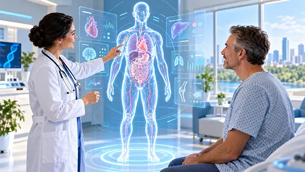
A digital twin of a human being sounds like a complete virtual copy of a patient, but medicine in 2026 is both more practical and more cautious than that description suggests. The most advanced systems usually reproduce a particular organ, disease process or aspect of a person’s physiology rather than every process in the body. They combine information such as medical imaging, laboratory results, electronic health records, physiological measurements and, in some cases, data from wearable devices or genetic testing. Doctors and researchers can then use the resulting model to examine how a patient’s condition may change or how different interventions could affect it. Cardiology currently provides some of the clearest clinical examples, while diabetes management, cancer care and other specialties are developing their own approaches. These models are becoming more capable, but their usefulness depends on good patient data, careful validation and clear evidence that their predictions are reliable enough for the particular medical decision being made.
What a Human Digital Twin Actually Means in Medicine
A medical digital twin is more than a three-dimensional picture of an organ. A CT or MRI reconstruction can show the shape of a heart, blood vessel or tumour, but a digital twin attempts to represent behaviour as well as structure. For example, a model of a patient’s heart may reproduce how electrical signals move through healthy and damaged tissue, allowing specialists to test where an abnormal rhythm could begin. A metabolic model may use changing glucose and insulin information to estimate how a person’s body could react under different circumstances. What matters is that the model represents a particular real patient and can be refined as more information becomes available.
The US Food and Drug Administration describes a digital twin as a set of information constructs that mimics the structure, context and behaviour of a physical counterpart and is dynamically updated using information from that counterpart. In healthcare, that distinction is important. A general mathematical model of a disease can help researchers understand typical patterns, but a patient-specific twin attempts to adjust those patterns to the individual. Age, anatomy, previous treatment, laboratory measurements, imaging findings and other relevant characteristics may all influence the result. The amount of personalisation varies considerably between systems, which is one reason the term “digital twin” does not yet describe a single standardised medical product.
It is also important to separate current medical reality from the idea of an exact virtual human. In 2026, most practical digital twins remain focused on a limited clinical problem. Researchers can build detailed models of hearts, vessels, metabolic responses or selected disease pathways, but reproducing every interaction between the brain, immune system, hormones, organs, medicines, genetics, behaviour and environment is far more difficult. Recent scientific reviews describe existing systems as predominantly organ-specific or disease-specific, with many still at the research, prototype or early clinical-validation stage. A full-body twin that doctors can routinely consult throughout a person’s life therefore remains a longer-term objective rather than an established part of ordinary healthcare.
How Patient Data Becomes a Working Personal Model
The process normally starts with information already collected during medical care. Imaging can provide the anatomy of an organ, while blood tests, electrocardiograms, medical history, medication records and physiological measurements add information about how that organ or system is functioning. Depending on the clinical question, researchers may also use molecular information, genetic results, pathology, exercise measurements, continuous glucose readings or patient-reported symptoms. The aim is not to collect every possible data point. A useful twin needs the information that is relevant to the decision it is intended to support.
Software then combines those measurements with established knowledge about biology and disease. A heart model, for instance, can be adjusted to match the size, shape and damaged areas visible in an individual patient’s scan. Simulations can then reproduce possible electrical activity or mechanical behaviour inside that personalised anatomy. Instead of carrying out an intervention immediately, clinicians may first test possible approaches in the model. This creates a controlled way to compare scenarios that cannot simply be tried repeatedly on a real patient. The model does not know the future with certainty; rather, it estimates what may happen under clearly defined assumptions.
A genuine clinical twin should also be capable of changing when the patient changes. A new scan, blood result, treatment response or monitoring record may alter the model and therefore alter its predictions. This is one of the features that separates the digital-twin idea from a static risk calculator. In practice, however, the frequency of these updates varies. Some models are created mainly for a single procedure, while others are intended to follow a condition over a longer period. The doctor remains responsible for interpreting the information because a simulation can be wrong when the incoming data are incomplete, when the patient’s condition falls outside the situations in which the model was tested or when important biological factors are not represented.
Where Patient-Specific Digital Twins Are Being Used and Tested in 2026
Cardiology has produced one of the strongest clinical examples so far. In April 2026, the New England Journal of Medicine reported a feasibility study involving ten patients with ventricular tachycardia, a potentially dangerous abnormal heart rhythm. Researchers created individual digital models of the patients’ hearts and used them to identify areas that could sustain the abnormal electrical circuits before catheter ablation. Following the procedures, ventricular tachycardia could not be induced in any of the ten participants. At an average follow-up of 13 months, eight remained free from recurrence without antiarrhythmic drug therapy. The study was small and cannot establish effectiveness for every patient, but it demonstrated that patient-specific simulations can be incorporated into a real treatment workflow rather than remaining solely a laboratory experiment.
Diabetes is another area in which the idea is moving towards practical testing. Automated insulin delivery already relies on continuous glucose information and algorithms that adjust insulin administration. Researchers are now studying whether a personalised model of a user’s changing physiology can make such systems more adaptive. A randomised clinical trial published in npj Digital Medicine in 2025 tested digital-twin technology as part of a system designed to improve the interaction between people with diabetes and automated insulin delivery. By 2026, diabetes research includes a growing range of digital-twin approaches involving glucose behaviour, insulin response, lifestyle information and treatment simulation. Reviews of the field nevertheless note that many studies remain small or require longer validation before broad clinical adoption can be justified.
Oncology illustrates a different stage of development. Cancer treatment generates large amounts of information from imaging, pathology, genomics, medicines, treatment response and follow-up. Researchers are investigating whether these separate records can be combined into evolving patient models that represent both the cancer and the person’s changing health. In July 2026, researchers from the Medical University of Vienna and collaborating institutions described a Virtual Human Twin approach for breast cancer decision support, with high-risk triple-negative breast cancer proposed as an important use case. The objective is to compare plausible treatment trajectories and make uncertainty visible to clinicians. This remains a developing research direction rather than a routine replacement for multidisciplinary cancer teams.
Why Digital Heart Twins Are Among the Most Advanced Examples
The heart is particularly suitable for personalised modelling because several important aspects of its structure and activity can be measured directly. MRI and CT scans provide detailed anatomy, while electrocardiograms and other cardiac tests show electrical or mechanical behaviour. Decades of cardiovascular research have also produced mathematical descriptions of how electrical signals travel through cardiac tissue and how damaged areas can change those signals. When these sources are combined, researchers can create a model that resembles a specific patient’s heart closely enough to ask focused questions about rhythm, blood flow or treatment planning.
Ventricular tachycardia shows why this can matter clinically. The abnormal electrical pathway responsible for an arrhythmia may pass through or around scarred heart tissue, and identifying the critical area can be difficult during an invasive procedure. A patient-specific simulation offers another source of information before treatment begins. Doctors can reproduce possible electrical circuits virtually and identify tissue that appears important for maintaining the rhythm. The 2026 TWIN-VT feasibility study provided an early demonstration of this approach in real patients. Its encouraging results do not mean that digital-twin-guided ablation has become a universal standard of care; larger studies, comparisons with existing techniques and longer follow-up remain necessary.
Researchers are also learning how to produce heart models at much greater scale. A 2025 study generated cardiac digital-twin resources from MRI data involving roughly 55,000 UK Biobank participants. Work of this kind is different from creating a model for an individual procedure, but it helps researchers understand normal variation and disease patterns across large populations. That population knowledge may eventually make personal models easier to build and interpret. Similar work is being conducted for diabetic heart disease, coronary circulation, cardiomyopathies and other cardiovascular conditions. The strongest future systems are therefore likely to combine two levels of information: detailed measurements from one patient and evidence derived from many patients with comparable characteristics.
What Digital Twins Can and Cannot Do for Patients in 2026
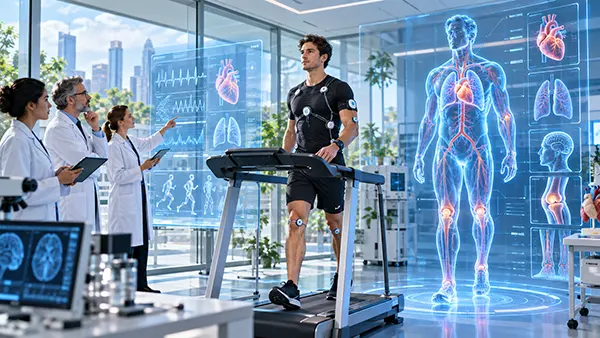
The clearest potential benefit of a patient-specific twin is the ability to compare options before making an irreversible decision. A surgeon or cardiologist may use a model to examine different intervention strategies, while researchers can simulate how a disease might respond under several treatment assumptions. Digital twins can also support what are often called in-silico studies, where some questions about medicines or medical devices are investigated using computational patients before or alongside conventional clinical research. The FDA specifically recognises potential uses for digital twins in personalised medicine and in simulated clinical studies. These applications can help narrow the range of options that require physical testing, but they do not remove the need for appropriate human clinical evidence.
Another advantage is the possibility of following change rather than relying on isolated snapshots. Traditional clinical decisions often depend on separate appointments, scans and laboratory results. A longitudinal twin could bring those observations together and show how the patient’s estimated state has altered over time. This is particularly relevant to chronic conditions such as diabetes, cardiovascular disease and cancer, where treatment decisions can change as the disease progresses or responds. The same principle could eventually support rehabilitation, treatment monitoring and prevention. The value comes from organising clinically meaningful information around an individual patient, not from producing a visually impressive virtual body.
There are equally important limits. A digital twin can only be as dependable as the measurements, medical knowledge and assumptions used to create it. Missing records, poor-quality scans, incorrect sensor readings or unrepresentative training data can all affect the result. A model tested mainly in one patient population may also perform less reliably in another. Systematic reviews published in 2026 continue to identify limited external validation, small study populations, inconsistent reporting and relatively little integration into routine clinical care. For patients, this means the label “digital twin” should never be treated as evidence that a tool has already been proven to improve diagnosis or treatment. The clinical claim made for each system has to be evaluated separately.
What Must Change Before Whole-Body Digital Twins Become Routine
Building a useful whole-person model requires far more than joining several organ models together. The body operates through constant interaction between cardiovascular, metabolic, nervous, immune and hormonal systems, while medicines, sleep, diet, physical activity, age and other factors can alter those relationships. Data are also collected at different times and for different purposes. A scan might be performed once a year, laboratory tests every few months and wearable measurements every few minutes. Connecting these sources without losing their medical meaning is a major challenge. Researchers therefore need common standards, reliable ways to exchange information and clear methods for showing how uncertain a prediction is.
Europe is making a substantial investment in this area through its Virtual Human Twins Initiative. The European Commission states that more than €100 million has been committed to work supporting virtual representations of cells, tissues, organs and organ systems, with applications including personalised care, clinical research, medical training and treatment planning. Work is also under way to improve access to modelling resources, high-performance computing and shared technical standards. The direction of travel is towards models that can connect several biological scales and eventually several organs, but European programmes themselves treat validation, privacy, interoperability and adoption by healthcare services as central tasks rather than solved problems.
The most realistic role for digital twins in the near future is therefore as decision-support tools used alongside clinicians, not autonomous substitutes for them. A trustworthy model should make clear what patient data it uses, what outcome it is estimating, how uncertain that estimate is and whether it has been validated in people similar to the patient in question. Doctors also need a way to recognise when the model is operating outside the circumstances for which it was tested. In 2026, digital twins are already moving beyond theory in selected areas, particularly personalised cardiac modelling, while wider applications in diabetes, oncology and multi-organ medicine continue to develop. Their progress will be measured less by how closely a computer image resembles a human body and more by whether carefully validated models help patients and clinicians make safer, better-informed decisions.